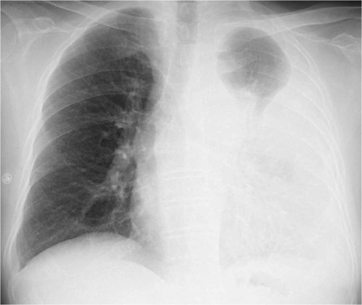
Loculated Pleural Effusion Diagram - Chest radiograph showing a left-sided, loculated pleural ... - Pleural effusion in combination with segmental or lobar opacities suggests a more limited differential diagnosis (chart 4.3).. Pleural effusions are abnormal accumulations of fluid within the pleural space. Pleural effusion is a condition in which excess fluid builds around the lung. Treatment depends on the cause. Case contributed by dr prashant mudgal. Is it localized to 1 specific area and does not move around when you.
Can someone clarify what a loculated pleural effusion is? The cause is sometimes respiratory, but there are several other. Pleural infection pleural inflammation pleural malignancy (most often pleural fluid analysis findings: They may result from a variety of pathological processes which overwhelm the pleura's ability to reabsorb fluid. More written notes and diagrams about pleural effusions are available on the website at www.zerotofinals.com/pleuraleffusion.
A) Loculated pleural effusion. A complex pleural effusion ... from www.researchgate.net Learn step 2 and shelf essentials in a free 10 min video. Bilateral pleural effusions withmeniscus signs. Pleural effusions can loculate as a result of adhesions. Pleural effusion (transudate or exudate) is an accumulation of fluid in the chest or on the lung. Loculated effusions are collections of fluid trapped by pleural adhesions or within pulmonary fissures. Other causes are complicated parapneumonic effusion. Treatment depends on the cause. Pleural effusion, or water on the lung, can resemble a respiratory infection.
Treatment depends on the cause.
Learn step 2 and shelf essentials in a free 10 min video. A pleural effusion is an accumulation of fluid within the pleural space. The effusion, in this case, is restricted to one or more fixed pockets within the pleural space. Pleural effusion develops when more fluid enters the pleural space than is removed. Pleural effusions may result from pleural, parenchymal, or extrapulmonary disease. Pleural effusion is the accumulation of fluid in the pleural space resulting from disruption of the homeostatic ct shows a loculated pleural fluid collection in association with pleural thickening and calcification. Other causes are complicated parapneumonic effusion. Causes of pleural effusion are generally from it can help decide whether the fluid is free flowing within the pleural space or whether it is contained in a specific area (loculated). When you have a pleural effusion, fluid builds up in the space between the layers of your pleura. Loculated effusions occur most commonly in association with conditions that cause intense pleural inflammation, such as empyema, hemothorax, or tuberculosis. Pleural effusions are abnormal accumulations of fluid within the pleural space. Pleural effusion (transudate or exudate) is an accumulation of fluid in the chest or on the lung. The cause is sometimes respiratory, but there are several other.
Thoracentesis is a simple bedside procedure with imaging guidance that permits fluid to be rapidly sampled, visualized, examined microscopically, and quantified for chemical and cellular content. An exudative pleural effusion occurs when there is increased permeability of the pleural surface and/or capillaries, usually as a result of inflammation. Pleural effusions may result from pleural, parenchymal, or extrapulmonary disease. 400+ pages of notes with diagrams, tables, tips and insight into topics. Empyema is defined as the presence of pus in the pleural space.
Peritoneal and meningeal relapse from lung adenocarcinoma ... from www.spandidos-publications.com The effusion, in this case, is restricted to one or more fixed pockets within the pleural space. Other causes are complicated parapneumonic effusion. Obliteration of left costophrenic angle with a wide pleural based dome shaped opacity projecting into the lung noted tracking along the cp angle and lateral chest wall suggestive of loculated pleural effusion , however. Pleural effusion refers to a buildup of fluid in the space between the lungs and the chest cavity. It can result from pneumonia and many other conditions. 138 097 просмотров 138 тыс. A pleural effusion is accumulation of excessive fluid in the pleural space, the potential space that surrounds each lung. Differentiation of loculated effusions from solid.
no change in position of effusion withchange in position of chest.
A pleural effusion is an accumulation of fluid within the pleural space. Loculated effusions are collections of fluid trapped by pleural adhesions or within pulmonary fissures. Parapneumonic effusion is a pleural fluid collection in association with an underlying pneumonia. Empyema is defined as the presence of pus in the pleural space. Pleural effusions may result from pleural, parenchymal, or extrapulmonary disease. Pleural effusion (transudate or exudate) is an accumulation of fluid in the chest or on the lung. Large right effusion (red arrow) displacesthe heart to the left (yellow arrow). 138 097 просмотров 138 тыс. Improved after thoracentesis and diuresis. Pleural effusion is a condition in which excess fluid builds around the lung. Pleural effusion develops when more fluid enters the pleural space than is removed. The pleura are thin membranes that line the lungs and the inside of the chest cavity and act to lubricate and facilitate breathing. The cause is sometimes respiratory, but there are several other.
Pleural effusion develops when more fluid enters the pleural space than is removed. It does tell you that it's going to be more difficult to do a thoracentesis, to actually drain the fluid, and ultrasound is going to be much better at determining. Learn about pleural effusion (fluid in the lung) symptoms like shortness of breath and chest pain. They may result from a variety of pathological processes which overwhelm the pleura's ability to reabsorb fluid. More written notes and diagrams about pleural effusions are available on the website at www.zerotofinals.com/pleuraleffusion.
(a) CXR-PA showing veiling opacity (resembling pneumonia ... from www.researchgate.net Terminology pleural effusion is commonly used as. Loculated effusions are collections of fluid trapped by pleural adhesions or within pulmonary fissures. Pleural infection pleural inflammation pleural malignancy (most often pleural fluid analysis findings: This is typically a chronic process. Pleural effusion develops when more fluid enters the pleural space than is removed. When you have a pleural effusion, fluid builds up in the space between the layers of your pleura. Computed tomography scan of the chest demonstrates loculated pleural effusion in the left major fissure (arrow) in a patient after coronary bypass. Thoracentesis is a simple bedside procedure with imaging guidance that permits fluid to be rapidly sampled, visualized, examined microscopically, and quantified for chemical and cellular content.
Ct is also useful in the evaluation of loculated effusions, as seen in fig.
Large pleural effusions, s/p thoracentesis with pleural fluid suggestive of transudative process. Thoracentesis is a simple bedside procedure with imaging guidance that permits fluid to be rapidly sampled, visualized, examined microscopically, and quantified for chemical and cellular content. Pleural effusions can loculate as a result of adhesions. Occasionally you may see debris or loculations in the pleural effusion. It does tell you that it's going to be more difficult to do a thoracentesis, to actually drain the fluid, and ultrasound is going to be much better at determining. Large right effusion (red arrow) displacesthe heart to the left (yellow arrow). Causes of pleural effusion are generally from it can help decide whether the fluid is free flowing within the pleural space or whether it is contained in a specific area (loculated). The cause is sometimes respiratory, but there are several other. no change in position of effusion withchange in position of chest. Bilateral pleural effusions withmeniscus signs. An exudative pleural effusion occurs when there is increased permeability of the pleural surface and/or capillaries, usually as a result of inflammation. Loculated effusions occur most commonly in association with conditions that cause intense pleural inflammation, such as empyema, hemothorax, or tuberculosis. Most likely secondary to left ventricular diastolic dysfunction.
Pleural effusion in combination with segmental or lobar opacities suggests a more limited differential diagnosis (chart 43) loculated pleural effusion. Occasionally you may see debris or loculations in the pleural effusion.


Posting Komentar
0 Komentar